- Branch Retinal Vein Occlusion
- Central Retinal Vein Occlusion
- Central Serous Chorioretinopathy
- Diabetic Retinopathy
- Epiretinal Membrane
- Lattice Degeneration
- Macular Hole
- Posterior Vitreous Detachment
- Retained Lens Fragments
- Retinal Artery Occlusion
- Retinal Detachment
- Retinal Tears
- Vitreomacular Traction
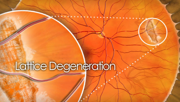
Lattice Degeneration
Clinically, lattice degeneration is characterized by oval or straight patches of thinned retina, sometimes accompanied by pigment clumps or a crosshatching pattern formed by sclerotic vessels (Figure 1). Lattice may be found in only one eye, but often is present in both. There may be just one lesion or clusters of many. Lattice is often located along the outer border of the retina.
Symptoms
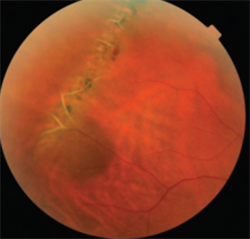
Hamid Ahmadieh, MD, Labbafinejad Medical Center. Retina Image Bank 2015; Image 25882. © 2018, American Society of Retina Specialists.
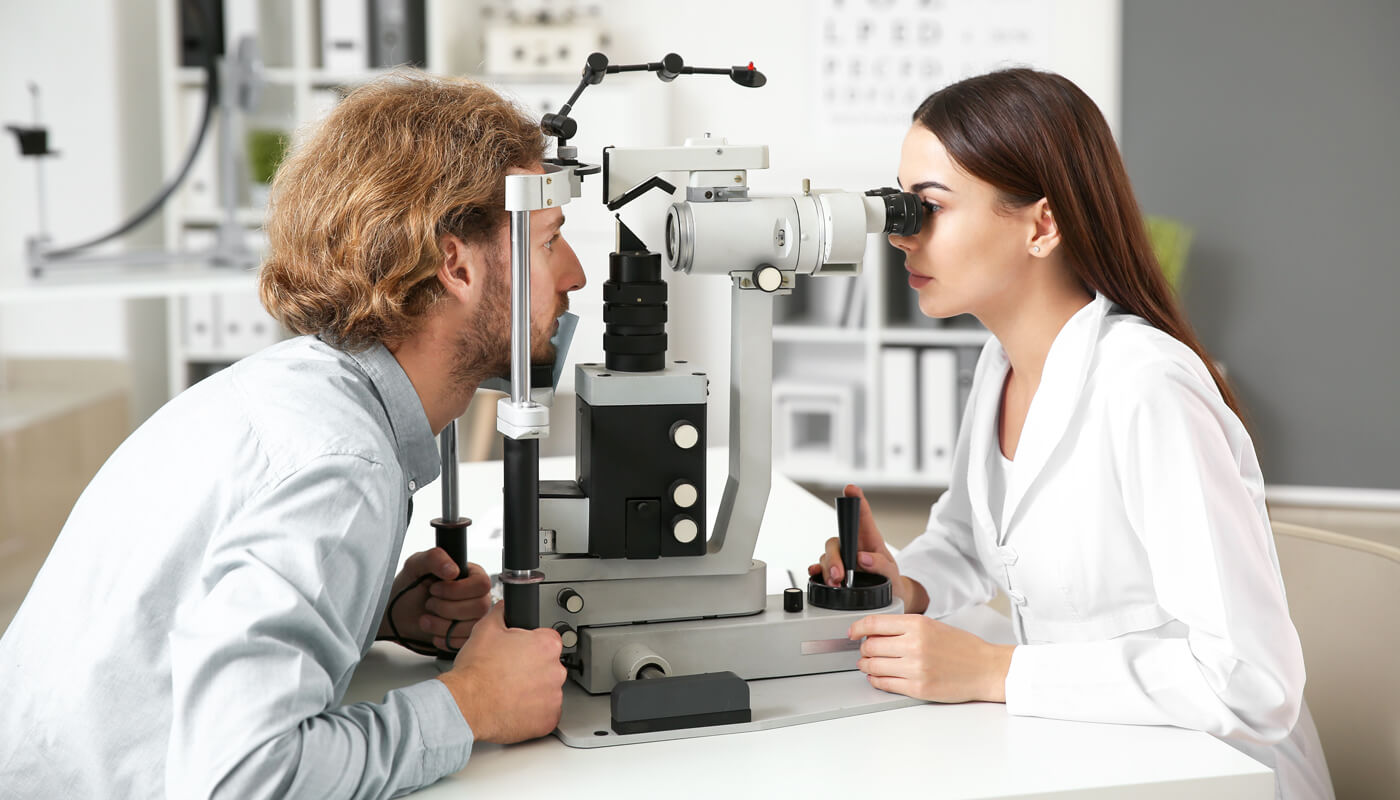
The eye care specialist will then use a headlight and a special lens to perform the exam. Depending on the nature of your findings, the doctor may also examine your retina while exerting a slight amount of pressure around your eye (scleral depression). In general, no imaging tests are necessary to diagnose this disease, although some providers may obtain wide-angle photographs of your retinas to assist in monitoring your condition.
- Quick diagnosis
- Complex medical tests
- Early identification and intervention
- Complex surgical interventions
Causes
Lattice degeneration occurs in 8% to 10% of the general population and its cause is not fully understood. There does not appear to be a correlation in incidence by gender or race. And while this condition does not follow a definitive inheritance pattern, it frequently clusters within families. It is most commonly found in patients with myopia
(nearsightedness), but lattice-like lesions are also seen in rare diseases such
as Stickler syndrome, Ehlers-Danlos, and Marfan syndrome. There is no
prevention or cure for lattice degeneration.
Risk factors
Symptoms include:
- Blurred vision
- Flashing lights
- Floaters
- Curtain obscuring part of your peripheral visual field
- Quick diagnosis
- Complex medical tests
- Early identification and intervention
- Complex surgical interventions
Treatment and Prognosis
In rare circumstances, some physicians may perform preventive laser therapy or cryotherapy (a type of freezing treatment) to strengthen the peripheral retina in the areas where it is weak. Your doctor will discuss the risks and benefits of doing so with you if he or she thinks it is worth considering. It is not known if these interventions are effective in preventing retinal complications.
Rarely, lattice degeneration can lead to complications such as a retinal tear or detachment. Symptoms to look out for include blurry vision, a curtain obscuring part of the outer visual field, flashing lights, and new floaters. If any of these occur, you should seek prompt attention from an ophthalmologist or retina specialist.
In those who do develop a retinal tear or detachment, treatment will be performed, typically by a retina specialist. This treatment can range from a laser treatment completed in the office to surgery in the operating room, depending on the severity of the condition.
In summary, lattice degeneration is a relatively common condition that affects the retina, especially in nearsighted people. While its presence increases the risk of a retinal tear or detachment, the vast majority of patients will never experience symptoms or complications from their lattice degeneration.
If you are diagnosed with lattice degeneration, the most important actions you can take to protect the health of your eyes are to visit your eye care provider on a regular basis and know the symptoms of a retinal tear or detachment so you can seek prompt treatment if needed.
Eye Procedures
Lasik Eye Surgery
- At vero eos et accusamus et iusto odio
- But I must explain to you how all this
- praising pain was born and I will
Contact Info
San Diego
7695 Cardinal Court, Suite 100 San Diego, CA 92123
Office: (858) 609-7100
Fax: (858) 609-7106
info@sdretina.com
Oceanside
3231 Waring Court, Suite S, Oceanside, CA 92056
Office: (760) 631-6144
Fax: (760) 724-3920
info@sdretina.com
Locations
San Diego
Oceanside
Get In Touch