- Branch Retinal Vein Occlusion
- Central Retinal Vein Occlusion
- Central Serous Chorioretinopathy
- Diabetic Retinopathy
- Epiretinal Membrane
- Lattice Degeneration
- Macular Hole
- Posterior Vitreous Detachment
- Retained Lens Fragments
- Retinal Artery Occlusion
- Retinal Detachment
- Retinal Tears
- Vitreomacular Traction

Epiretinal Membrane
Symptoms

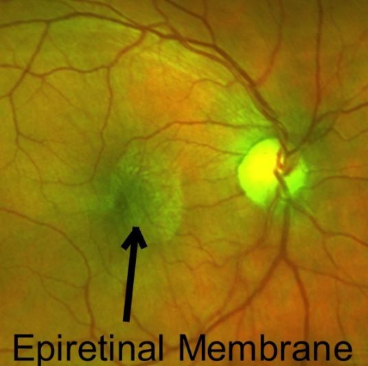
Patients may notice metamorphopsia, a symptom that causes visual distortion in which shapes that are normally straight, like window blinds or a door frame, looking “wavy” or “crooked,” especially when compared to the other eye. In advanced cases, this can lead to severely decreased vision. Less commonly, ERMs may also be associated with double vision, light sensitivity or images looking larger or smaller than they actually are.
Causes
The most common cause of macular pucker is an age-related condition called posterior vitreous detachment (PVD), where the vitreous gel that fills the eye separates from the retina, causing micro-tears and symptoms of floaters and flashes. If there is no specific cause apart from the PVD, the ERM is called idiopathic (of unknown origin.)
ERMs can be associated with a number of ocular conditions such as prior retinal tears or detachment, retinal vascular diseases such as diabetic retinopathy or venous occlusive disease; they can also be post-traumatic, occurring following ocular surgery, or be associated with intraocular (inside the eye) inflammation.
Risk factors
- Quick diagnosis
- Complex medical tests
- Early identification and intervention
- Complex surgical interventions
Diagnostic testing
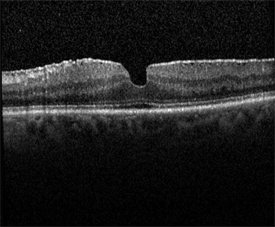
Most ERM cases can be diagnosed by an eye care provider during a routine clinical exam. Ocular Coherence Tomography (OCT) is an important imaging method used to assess the severity of the ERM (Figure 1). Sometimes, additional testing such as fluorescein angiography is used to determine if other underlying retinal problems have caused the ERM.

Sharon Fekrat, MD, FACS, Duke University Eye Center. Retina Image Bank 2012; Image 1437. ©American Society of Retina Specialists.
- Quick diagnosis
- Complex medical tests
- Early identification and intervention
- Complex surgical interventions
Treatment and Prognosis
Since most ERMs are fairly stable after an initial period of growth, they can simply be monitored as long as they are not affecting vision significantly. In rare circumstances, the membrane will spontaneously release from the retina, relieving the traction and clearing up the vision. However, if an exam shows progression and/or functional worsening in vision, surgical intervention may be recommended.
There are no eye drops, medications or nutritional supplements to treat ERMs. A surgical procedure called vitrectomy is the only option in eyes that require treatment. With vitrectomy, small incisions are placed in the white part of the eye, and the vitreous gel filling the inside of the eye is replaced with saline. This allows access to the surface of the retina where the ERM can be removed with delicate forceps, thereby allowing the macula to relax and become less wrinkled.
The risk of complications with vitrectomy is small, with about 1 in 100 patients developing retinal detachment and 1 in 2000 developing infection after surgery. Patients who still have their natural lens will develop increased progression of a cataract in the surgical eye following surgery.
Factors affecting visual outcome include:
- Length of time the ERM has been present
- The degree of traction (or pulling)
- The cause of the ERM (Idiopathic ERMs have a better prognosis than eyes with prior retinal detachment or retinal vascular diseases
Surgery for ERM has a good success rate, and most patients experience improved visual acuity and decreased metamorphopsia following vitrectomy.
Eye Procedures


Lasik Eye Surgery
- At vero eos et accusamus et iusto odio
- But I must explain to you how all this
- praising pain was born and I will
Contact Info
San Diego
7695 Cardinal Court, Suite 100 San Diego, CA 92123
Office: (858) 609-7100
Fax: (858) 609-7106
info@sdretina.com
Oceanside
3231 Waring Court, Suite S, Oceanside, CA 92056
Office: (760) 631-6144
Fax: (760) 724-3920
info@sdretina.com
Locations
San Diego
Oceanside
Get In Touch

